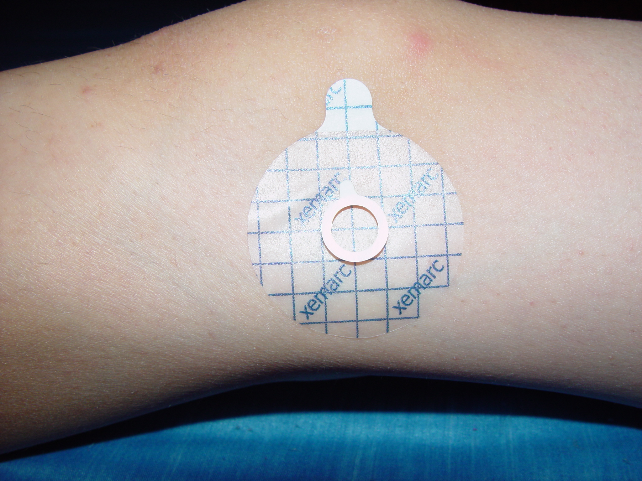
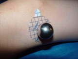
Placement of MCM
The MCM not only needs to be on the image at acquisition, it needs to be placed at bone level of the area of interest or planning.
For example: If the patient is having a pre-operative hip x-ray and the views are an AP Pelvis, AP Hip and a Lateral Hip (shoot-thru or frogged), the sphere should be placed on all images:
AP Pelvis: Placed at the level of the greater trochanter. Optimal placement is medially, midline to femurs and proximally towards the symphysis. Lateral placement is OK if the patient doesnt have a large amount of adipose tissue. If the patient is larger, placement will need to be carefully placed medially.
AP Hip: Same placement as above
Lateral Hip view (frogged): Place the MCM laterally at the level of the greater trochanter.
Lateral Hip view (Shoot-thru): Place the MCM anteriorly, at the level of the greater trochanter.
Creative placement is sometimes required, x-ray technologists are required to be creative in their job every day!
In the example of a full-leg that is being acquired for knee joint axis correction, place the sphere preferably medially but can be laterally at the level of the knee joint.
* Please ensure that enough MCM's are ordered to be kept in each room in which Orthopaedic cases will be acquired.
* It is up to the individual radiological sites to determine a protocol of MCM placement between Radiology and the Orthopaedic Surgeons.
Warning: You can only scale the image correctly if a scaling MCM was placed in the image area while the image was exposed. Check your measurements carefully. False values can have serious consequences for the patient regarding misinterpretations of the right prostheses size.